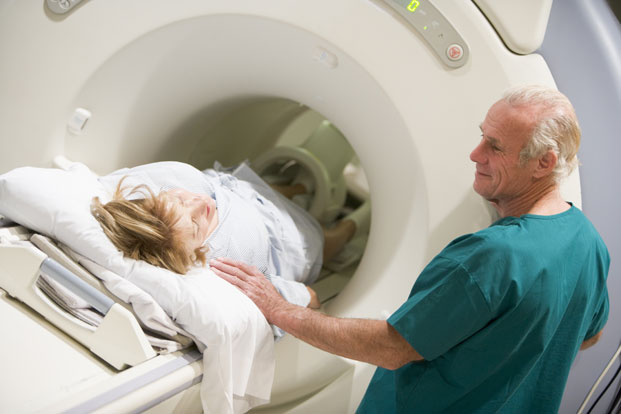
There’s controversy in some parts of the medical world which concerns the use of X-rays and CT scans. Use of radiation as a medical imaging tool has been around for about 120 years. Computed tomography, or CT, has been around since the 1970s. Use of the CT scanner has risen dramatically over the past 40 years. In 1980, there were 3 million scans done. That number now hovers around 80 million. The issue centers around the levels of radiation patients are exposed to while undergoing diagnostic tests.
What is radiation?
Radiation is a form of energy, particle waves moving through space. Typically, most of the radiation we constantly receive from the environment is through sun exposure, radon gas, and cosmic rays from space. This is called “background” radiation, and the higher your altitude, the more powerful the background radiation to which you are exposed. But studies show that nowadays, we get half our lifetime radiation levels from medical imaging devices.
X-rays and CT scans use “ionizing” radiation. This radiation type is considered unstable because it has excess energy it must get rid of. This unstableness is what makes a substance radioactive. These X-rays penetrate the body, straight through human tissue to see broken bones, swallowed objects and cavities. There is a modified X-ray technique that allows the imaging of softer tissues like blood vessels, intestines and lungs.
How CT Scans Work
Computerized axial tomography (CAT) is another name for CT scans as they accomplish the same thing in the same manner. As we noted, X-rays are everywhere, but these rays are powerful examples of electromagnetic energy focused to do a job.
X-ray beams move around the person, who is typically lying on a table. The machine shoots horizontal X-ray pictures of the subject from many different angles. Each full rotation around the body results in a scan of a slice of the body. A computer then takes the information of multiple scans and puts together a 3D image of that body section.
CT scans can emit radiation doses equivalent to 1,000 to 2,000 chest X-rays, more or less, depending on the size of the scanned body section. This is about seven years of natural sourced exposure for humans. And it is the use of this level of radiation that concerns some researchers and physicians.
DNA, anyone?
You knew we had to address this issue. Radiation has long been known as a carcinogenic force, but how does it do it? Because of radiation’s unstable nature and its ability to penetrate the human body, radiation can cause “free radicals.” These are unstable cells that rob healthy ones of a component, thus making unstable, unhealthy cells out of them.
These cells don’t act as normal cells because their DNA, the blueprint of the cell and its functions, is damaged. These cells don’t die off as normal cells would, and they can invade other tissues, something healthy cells cannot do. Because they don’t die off, masses of these DNA-damaged cells may accumulate as cancerous tumors.
The Controversy
This is where the “he said, she said” part comes in because there haven’t been enough studies on the effects of powerful X-rays on the human body. And because of that, you have two sides to the medical saga of radiation usage.
In a 2005 report, the National Academy of Sciences stated that there wasn’t a radiation threshold, below of which could be deemed harmless. In other words, no radiation is good radiation. But because that is impossible in these days of modern medical science and shrinking ozone layers, the few studies that exist show a low risk, but a risk, of radiation-induced cancers.
To illustrate, a CT scan of a chest is equal to up to 2,000 chest X-rays in radiation exposure numbers, depending on area size. An abdominal scan, a smaller area, could equal 500 chest X-rays.
Researchers estimate that two percent of future cancers in the U.S. could be attributable to medical radiation uses, including CT and CAT scans. That comes out to be equal to about 29,000 cases of cancer a year with 15,000 deaths.
A 2013 Australian study determined that overall, people who had CT scans had a 24 percent higher chance of getting cancer than someone who hadn’t undergone the procedure. Each additional scan, it was found, added another 16 percent of acquiring cancer because of these imaging techniques.
Though a CT holds a cancer risk of possibly fatal illnesses as one chance in 2,000, the overall risk of cancer in the U.S. population is one in five. Ha!, say supporters, the risk of a CT scan is miniscule. In a situation where the diagnosis of an illness outweighs the benefits of not getting a scan, a scan is warranted.
When to Ask Questions
If a doctor would like you to have a CAT or CT scan, it is time to ask questions. First, ask why the imaging test and if another diagnostic test could be used instead, like an ultrasound or MRI, both of which don’t use radiation to do their jobs.
Because there are no federal limits on CT scans, keep records for yourself. Some doctors may not be aware of how much radiation to which you have already been exposed.
And beware the uninformed physician. A 2012 survey of a population of doctors revealed that half did not realize that CT scans increase the patient’s risk of cancer. Another report estimated about 35 percent of CTs were ordered as a defense against medical lawsuits. These “just-in-case” exams add to the risk of radiation-induced cancers like bone, leukemia, thyroid, breast, lung and skin cancers, which could take decades to develop. So do your homework and don’t be afraid to question your doctor as to his or her treatment protocol. It is, after all, your health.

Leave a Reply